 dorris
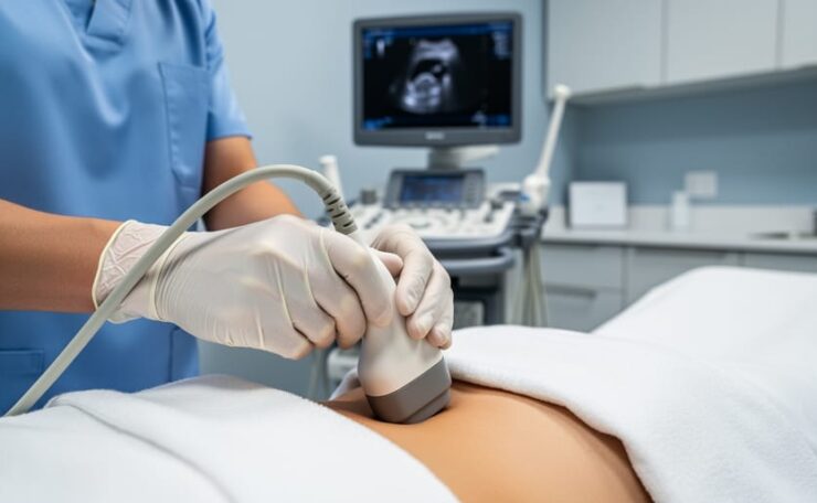
Yes, endometriosis can be seen on ultrasound, but with important limitations. Transvaginal ultrasound is quite sensitive at detecting ovarian endometriomas (those characteristic “chocolate cysts”) and can identify deep infiltrating endometriosis when performed by an experienced examiner. However, it cannot reliably detect superficial peritoneal endometriosis, which is one of the most common forms of the disease.
If you’ve been experiencing pelvic pain, painful periods, or fertility challenges and your doctor has ordered an ultrasound, you’re likely wondering whether this test will finally provide answers. The truth is more nuanced than a simple yes or no. While ultrasound has become an increasingly valuable tool in the endometriosis diagnostic journey, what it reveals depends on the type and location of endometriosis tissue.
I understand how frustrating it can be to undergo testing without knowing what to expect or whether you’ll walk away with a clear diagnosis. Many women spend years searching for answers, and knowing the capabilities and limitations of different diagnostic tools is essential for advocating for your own health care.
Recent research involving over 1,100 patients has demonstrated that transvaginal ultrasound can accurately assess the presence and extent of certain types of endometriosis. The key factor? Examiner experience matters tremendously, especially when diagnosing deep infiltrating endometriosis. This means the skill of the person performing your ultrasound can significantly impact what gets detected.
In this article, we’ll walk through exactly what ultrasound can and cannot see, how accuracy varies by endometriosis type, and what steps you might need to take if your ultrasound doesn’t provide the complete picture you’re hoping for.
Understanding the Different Types of Endometriosis and What Ultrasound Can See
Endometriosis isn’t one uniform condition. It exists in three distinct forms, and understanding these differences explains why your ultrasound might catch some signs while missing others entirely.
The first type, superficial peritoneal lesions, appears as shallow patches of endometrial-like tissue scattered across the lining of your pelvis and abdomen. These lesions sit on the surface of organs rather than burrowing into them. They’re the most common form, yet they’re essentially invisible on ultrasound. The tissue is too thin and flat to create the contrast needed for imaging to pick it up.
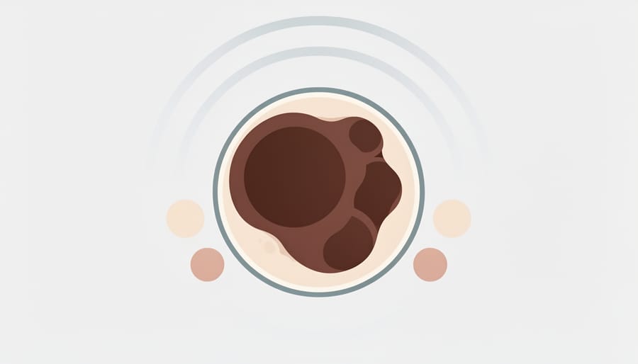
Ovarian endometriomas, commonly called chocolate cysts, are easier to spot. These fluid-filled cysts develop when endometrial tissue grows inside the ovary, creating a distinct appearance on imaging. The old blood trapped inside gives them a characteristic dark, homogeneous look that experienced sonographers recognize immediately. Transvaginal sonography is a quite sensitive tool for detecting these ovarian endometriomas, which is why they’re often the first sign of endometriosis that shows up on imaging.
The third type, deep infiltrating endometriosis, occurs when tissue burrows more than five millimeters beneath the surface of organs. This version commonly affects the bowel, bladder, and the space between the uterus and rectum. Because DIE creates thicker, more substantial masses, skilled examiners can often identify it on transvaginal ultrasound. The tissue’s depth and density create enough contrast for imaging to capture, though accuracy depends heavily on the sonographer’s experience with endometriosis specifically.
Location and depth determine visibility. Shallow surface lesions blend into surrounding tissue on ultrasound, while deeper growths and contained cysts create the shadows and distinct shapes that imaging can distinguish. This explains why two women with equally severe symptoms might have completely different ultrasound results.
How Transvaginal Ultrasound Works for Endometriosis Detection
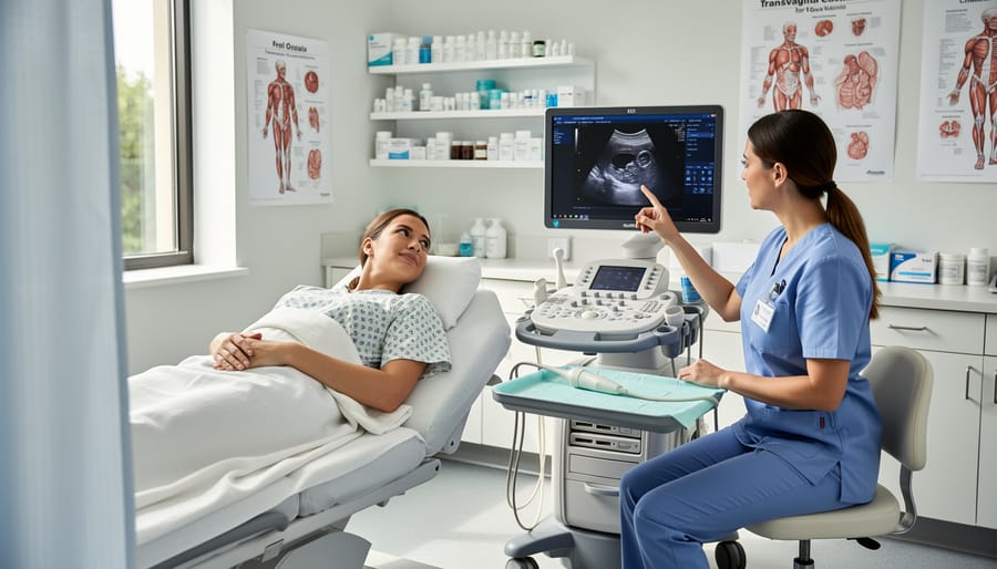
A transvaginal ultrasound involves placing a slim, wand-shaped probe into the vagina to capture detailed images of your pelvic organs. The probe sits much closer to your uterus and ovaries than an abdominal ultrasound can reach, which makes it far more effective at spotting endometriosis-related changes.
The exam typically takes 15 to 30 minutes. You’ll lie on your back with your knees bent, similar to a pelvic exam position. The sonographer applies gel to the probe, inserts it gently, and moves it to view different angles of your pelvis. Most women find the procedure manageable, though if you have pain from endometriosis, some pressure during the exam might feel uncomfortable. You can ask the sonographer to pause or adjust if needed.
What makes transvaginal sonography particularly valuable is its ability to detect ovarian endometriomas, those blood-filled cysts often called chocolate cysts because of their appearance. Research shows that TVS is a quite sensitive tool for detecting these ovarian lesions. The sonographer looks for specific characteristics: cysts with a ground-glass appearance, thick walls, and internal echoes that indicate old blood.
This method also works well for spotting deep infiltrating endometriosis, especially when it affects areas like the bowel, bladder, or the space between your vagina and rectum. The sonographer examines tissue thickness, nodules, and whether organs move freely or appear stuck together.
However, the accuracy of your scan depends heavily on the examiner’s experience with endometriosis. Studies across 1,119 patients have demonstrated varying sensitivity and specificity levels, with examiner skill being the most crucial factor for detecting deep infiltrating disease. Don’t hesitate to ask whether your sonographer regularly evaluates patients for endometriosis.
The Accuracy of Ultrasound for Endometriosis: What the Research Shows
When you’re trying to understand whether ultrasound can reliably detect endometriosis, you’re likely wondering: how accurate is this test, really? The research gives us some concrete answers, though they come with important nuances.
A comprehensive meta-analysis examining eight studies with 1,119 patients evaluated the accuracy of transvaginal ultrasound for detecting bowel endometriosis specifically. The study looked at two key measures: sensitivity (how good the test is at identifying endometriosis when it’s actually present) and specificity (how good it is at ruling it out when it’s not there). Think of sensitivity as the test’s ability to catch what’s there, while specificity is its ability to avoid false alarms.
The results showed that transvaginal ultrasound performs quite well for certain types of endometriosis, particularly ovarian endometriomas and deep infiltrating endometriosis in specific locations. For ovarian cysts caused by endometriosis, the sensitivity is notably high, meaning the test catches most cases. Deep infiltrating endometriosis affecting the bowel also shows up reasonably well on ultrasound when performed by experienced practitioners.
Here’s where it gets critical: accuracy varies dramatically based on both the location of endometriosis and who’s performing the scan.
A sonographer who specializes in endometriosis detection will identify far more cases than someone performing routine pelvic ultrasounds. Studies have also investigated how factors like age and body mass index affect diagnostic accuracy, finding that these variables can influence how clearly endometriosis appears on imaging.
What this means for you: a negative ultrasound doesn’t definitively rule out endometriosis, especially if the person performing your scan doesn’t specialize in detecting it. The test’s reliability depends heavily on technical expertise, the type of endometriosis present, and its location. This isn’t about the technology failing, it’s about the complexity of what we’re trying to see and the skill required to recognize it.
What Ultrasound Misses: The Limitations You Need to Know
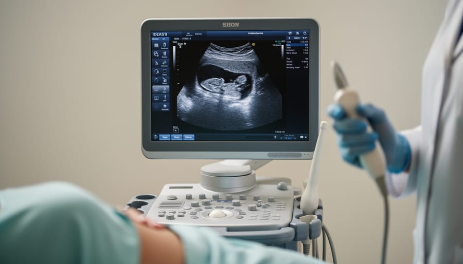
The most challenging limitation of ultrasound is its inability to detect peritoneal endometriosis, the superficial lesions that grow on the lining of the pelvic cavity. These shallow implants are often too thin to create distinct changes on imaging. They don’t form masses or alter tissue density enough to stand out from surrounding structures, which means they remain invisible even on high-quality transvaginal scans.
This creates a frustrating reality for many women. You might experience debilitating pain, heavy bleeding, and digestive issues that disrupt your daily life, yet your ultrasound comes back showing nothing abnormal. It’s an experience that leaves many feeling dismissed or wondering if they’re imagining their symptoms.
You’re not imagining anything. Normal imaging doesn’t mean normal health. Research confirms that compared to laparoscopy, transvaginal ultrasound has no diagnostic value for peritoneal endometriosis, meaning this common form of the disease simply cannot be seen through this method. The absence of visible findings reflects the technology’s limitations, not the absence of disease.
Beyond peritoneal lesions, ultrasound can also miss small areas of deep infiltrating endometriosis, particularly if the examining physician lacks specialized training in endometriosis imaging. The quality of your scan depends heavily on examiner experience, which varies widely among facilities.
If your ultrasound is normal but your symptoms persist, trust yourself. Document your symptoms in detail, including their timing, severity, and impact on your life. Seek evaluation from a gynecologist or specialist with specific endometriosis expertise. They may recommend additional imaging like MRI or refer you for further assessment. Your pain deserves investigation, regardless of what initial imaging shows.
Other Imaging Options and When They’re Used
While transvaginal ultrasound serves as the primary imaging method for endometriosis, other imaging tools can provide crucial information when evaluating disease extent, particularly for complex cases.
MRI stands out as a powerful complementary option. This imaging technique excels at mapping deep infiltrating endometriosis, especially when it involves the bowel, bladder, or structures deep in the pelvis. Your doctor might recommend MRI if your transvaginal ultrasound suggests extensive disease or if they need clearer visualization before planning treatment. The detailed images help surgeons understand exactly what they’ll encounter, which is invaluable for understanding conditions that require surgical intervention.
Transrectal ultrasound offers another perspective, particularly for assessing bowel involvement. This specialized technique provides detailed views of the rectum and lower bowel, areas where deep endometriosis commonly occurs. It’s typically reserved for cases where bowel endometriosis is suspected based on symptoms or findings from other imaging.
Here’s what matters for your care: reliable imaging contributes to accurate assessment of endometriosis presence and extent, which directly impacts treatment planning. Gone are the days when laparoscopy was the only definitive diagnostic option. Current guidelines recognize that laparoscopy cannot be considered purely a diagnostic tool, acknowledging that skilled imaging can often provide the information needed without surgery.
Your healthcare team will determine which imaging approach makes sense for your specific situation based on your symptoms, initial ultrasound findings, and treatment goals.
Real Women’s Experiences: Navigating the Diagnostic Journey
The diagnostic experiences of women with endometriosis vary widely, and understanding these patterns can help you navigate your own journey with greater confidence.
Some women receive clear answers through ultrasound. One representative scenario involves a woman experiencing severe pain and bloating who underwent transvaginal ultrasound that revealed a 5cm ovarian endometrioma. The visible cyst explained her symptoms and allowed her care team to develop a treatment plan immediately. She found relief in finally having a concrete explanation after years of being told her pain was “just bad periods.”
Others face a more complicated path. Another common experience involves women with debilitating symptoms, painful periods, digestive issues, fatigue, whose ultrasounds come back normal. In these cases, the peritoneal endometriosis causing their symptoms simply isn’t visible on imaging. These women often feel dismissed or confused, wondering if their pain is real when the scan shows nothing. Understanding that normal imaging doesn’t rule out endometriosis becomes crucial for these patients.
The empowerment comes from knowing what questions to ask and what to expect. Women who understand your diagnosis process, including ultrasound’s strengths and limitations, are better equipped to advocate for themselves. They know to seek experienced examiners for deep infiltrating disease, to document their symptoms carefully, and to persist when initial imaging doesn’t provide answers. This knowledge transforms the diagnostic journey from a confusing series of tests into a process where you remain an informed, active participant in your care.
What to Do If Your Ultrasound Results Don’t Match Your Symptoms
A normal ultrasound doesn’t mean your pain and symptoms aren’t real. If your imaging comes back clear but you’re still experiencing heavy periods, pelvic pain, or pain during sex, trust yourself and keep pushing for answers.
Start by keeping a detailed symptom diary. Note when pain occurs, its severity on a scale of one to ten, what triggers it, and how it affects your daily life. Track your menstrual cycle, bowel and bladder symptoms, and any patterns you notice. This documentation becomes powerful evidence when advocating for further investigation.
Your next step is finding a healthcare provider who specializes in endometriosis. General gynecologists may not have the expertise to recognize subtle presentations or know when additional evaluation is warranted. A specialist understands that peritoneal endometriosis won’t show on ultrasound and knows how to interpret your symptoms in context.
Ask specifically about MRI or getting a second ultrasound with a sonographer experienced in detecting deep infiltrating endometriosis. Examiner experience is the most crucial factor in ultrasound accuracy for complex cases. Sometimes a different imaging approach or a more skilled eye can reveal what was initially missed.
Remember that learning to pay attention to symptoms applies across all aspects of women’s health. Your body is telling you something, and normal imaging is just one piece of the puzzle, not the final answer. You deserve a provider who listens and continues investigating until you have real answers and relief.
Questions to Ask Your Healthcare Provider About Ultrasound and Endometriosis
Walking into a doctor’s appointment prepared with the right questions transforms you from passive patient to active participant in your care. When it comes to ultrasound for endometriosis, knowing what to ask helps you understand what the imaging can realistically reveal and what your next steps should be.
Start by asking about the sonographer’s specific experience with endometriosis imaging. Since examiner experience is the most crucial factor when diagnosing deep infiltrating endometriosis, you want to know how many endometriosis cases they’ve evaluated. A general sonographer and one who specializes in gynecological conditions will have vastly different detection rates.
Ask directly which types of endometriosis the ultrasound can detect. Request clarification about whether transvaginal sonography can identify ovarian endometriomas and deep infiltrating disease in your case, and acknowledge upfront that peritoneal endometriosis won’t be visible on this imaging.
What happens if my ultrasound looks normal but I still have symptoms?
A normal ultrasound doesn’t rule out endometriosis, particularly peritoneal disease which doesn’t show up on imaging. Your symptoms remain valid and warrant further evaluation with an endometriosis specialist.
Should I get an MRI instead of an ultrasound?
MRI can evaluate disease extent in complex cases, but transvaginal ultrasound is typically the first-line imaging. Your doctor will recommend MRI if they need more detailed mapping of deep infiltrating endometriosis.
How accurate is ultrasound for detecting bowel endometriosis?
Research involving 1,119 patients across eight studies shows transvaginal ultrasound has measurable accuracy for bowel endometriosis, though examiner experience significantly impacts results.
If results come back inconclusive, ask about next steps. Will they recommend specialist evaluation, additional imaging like MRI or transrectal ultrasound, or symptom management while monitoring? Understanding the plan removes uncertainty and empowers you to move forward with confidence. This kind of women’s health awareness ensures you receive thorough care that addresses your specific situation rather than accepting a one-size-fits-all approach.
Ultrasound remains one of the most valuable first-line tools we have for diagnosing certain types of endometriosis, particularly ovarian endometriomas and deep infiltrating disease. It’s accessible, non-invasive, and when performed by experienced practitioners, remarkably effective. But it’s not a complete solution. The reality that peritoneal endometriosis remains invisible on imaging means that a normal ultrasound doesn’t close the door on diagnosis or invalidate your symptoms.
Understanding what ultrasound can and cannot see puts you in a stronger position to advocate for yourself. If your symptoms persist despite normal imaging, that information itself is meaningful. It tells you and your healthcare team that further evaluation may be needed, whether that’s a different imaging approach, consultation with an endometriosis specialist, or a comprehensive symptom assessment.
Your health journey is yours to navigate, and being informed about the tools available makes you a more effective partner in your care. When we understand both the power and the limitations of diagnostic imaging, we can ask better questions, make more informed decisions, and persist when initial results don’t provide answers. That persistence matters, because you deserve thorough investigation and real solutions.